Contents
- ✨ What is Radiography?
- 🏥 Medical Applications: A Diagnostic Powerhouse
- 🏭 Industrial Radiography: Beyond the Body
- ✈️ Security Screening: A Glimpse of the Invisible
- 💡 How it Works: The Science of Seeing
- 🆚 Radiography vs. Other Imaging Modalities
- 💰 Cost & Accessibility: Navigating the System
- 🚀 The Future of Radiography: Evolution and Innovation
- Frequently Asked Questions
- Related Topics
Overview
Radiography is the foundational imaging science that allows us to peer inside objects, whether it's the intricate structure of the human body or the internal integrity of a manufactured component. At its heart, it employs various forms of radiation, most famously X-ray radiation, to create visual representations of internal structures. This technique is indispensable across multiple fields, from diagnosing ailments in medicine to ensuring quality control in industry and enhancing security at airports. The ability to visualize the unseen has fundamentally reshaped how we understand and interact with the physical world.
🏥 Medical Applications: A Diagnostic Powerhouse
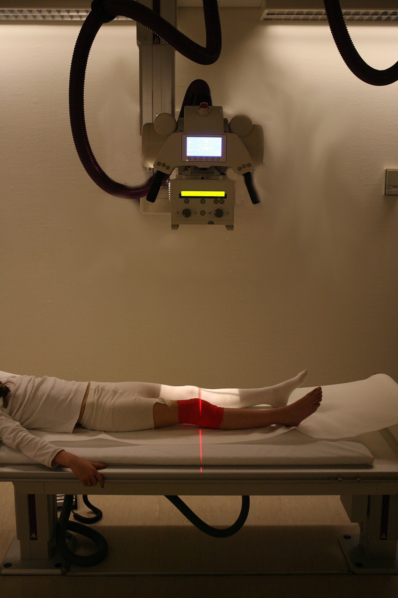
In the medical realm, radiography is nothing short of a diagnostic cornerstone. It's the go-to for detecting fractures in bones, identifying pulmonary issues like pneumonia, and locating foreign objects. Techniques like standard X-rays provide quick, two-dimensional snapshots, while CT scans offer cross-sectional views, allowing for detailed examination of soft tissues, organs, and blood vessels. The information gleaned from radiographic images directly informs treatment plans, making it a critical tool for diagnosing diseases and injuries.
🏭 Industrial Radiography: Beyond the Body
Beyond healthcare, industrial radiography plays a vital role in ensuring the safety and reliability of manufactured goods. It's used to inspect welds, detect internal flaws in castings, and examine pipelines for corrosion or cracks without damaging the material. This non-destructive testing (NDT) method is crucial in sectors like aerospace, automotive, and construction, where structural integrity is paramount. The principles are similar to medical radiography, but the focus shifts to material composition and structural defects rather than biological anatomy.
✈️ Security Screening: A Glimpse of the Invisible
At airports and other high-security checkpoints, radiography is a silent guardian. X-ray scanners are used to examine the contents of luggage and cargo, identifying prohibited items, weapons, or explosives. These systems are designed for rapid throughput, using specialized X-ray technology to differentiate between various materials based on their density and atomic number. While the underlying physics is shared with medical radiography, the engineering and operational requirements are tailored for security applications.
💡 How it Works: The Science of Seeing
The fundamental principle of radiography involves passing a beam of radiation through an object and detecting what emerges on the other side. Different materials absorb radiation to varying degrees based on their density and composition. Dense materials, like bone or metal, absorb more radiation, appearing lighter on the resulting image, while less dense materials, like soft tissue or air, allow more radiation to pass through, appearing darker. X-ray sources produce the initial beam, and detectors capture the attenuated radiation to form the image.
🆚 Radiography vs. Other Imaging Modalities
Radiography, particularly standard X-rays, is often the first-line imaging modality due to its speed and cost-effectiveness. However, it has limitations in visualizing soft tissues compared to MRI, which uses magnetic fields and radio waves. Ultrasound, employing sound waves, is excellent for real-time imaging of moving structures like fetuses or blood flow, and it avoids ionizing radiation altogether. CT scans offer superior detail for cross-sectional anatomy but involve higher radiation doses than plain X-rays.
🚀 The Future of Radiography: Evolution and Innovation
The field of radiography is not static; it's constantly evolving. Innovations in detector technology are leading to digital systems that offer higher image quality with lower radiation doses. Artificial intelligence (AI) is increasingly being integrated to assist radiologists in image interpretation, potentially speeding up diagnosis and improving accuracy. Furthermore, advancements in DXA scanning and spectral imaging are providing richer diagnostic information. The ongoing quest is for safer, faster, and more informative imaging solutions.
Key Facts
- Year
- 1895
- Origin
- Wilhelm Conrad Röntgen
- Category
- Medical Technology
- Type
- Technology
Frequently Asked Questions
Is radiography safe?
Medical radiography uses ionizing radiation, which carries a small risk of cellular damage. However, the benefits of accurate diagnosis typically far outweigh the risks, especially when doses are kept as low as reasonably achievable (ALARA). Regulatory bodies set strict guidelines for radiation exposure. Safety protocols are paramount in all radiography settings, both medical and industrial.
What's the difference between X-rays and CT scans?
Standard X-ray imaging produces a single, flat, two-dimensional image of the area of interest. CT scans, on the other hand, use X-rays to acquire multiple cross-sectional images (slices) as the source and detector rotate around the patient. These slices can then be reconstructed by a computer to create detailed 3D images, offering a more comprehensive view of internal structures.
How long does a typical radiography procedure take?
The duration varies greatly. A simple plain X-ray might take only a few minutes, including patient positioning. A CT imaging procedure, while the scanning itself is fast (often less than a minute), can take longer when accounting for patient preparation, setup, and image reconstruction. Contrast agent administration can also add time.
Do I need a referral for radiography?
In most healthcare systems, a referral or prescription from a qualified healthcare professional, such as a doctor or specialist, is required for radiographic examinations. This ensures the imaging is medically necessary and appropriate for your condition. Some direct-access screening programs may exist for specific tests.
What is the role of a radiographer?
A radiologic technologist is a trained healthcare professional who operates the radiographic equipment and positions patients to obtain diagnostic images. They ensure patient safety, manage radiation exposure, and produce high-quality images for interpretation by a radiologist. Radiographers are essential members of the diagnostic imaging team.